Can The Image Be Viewed Directly In A Light Microscope
Light microscope images can be viewed directly. The light microscope is an important tool in the study of microorganisms particularly for identification purposes.
 Differences Between Light Microscope And Electron Microscope
can the image be viewed directly in a light microscope
can the image be viewed directly in a light microscope is a summary of the best information with HD images sourced from all the most popular websites in the world. You can access all contents by clicking the download button. If want a higher resolution you can find it on Google Images.
Note: Copyright of all images in can the image be viewed directly in a light microscope content depends on the source site. We hope you do not use it for commercial purposes.
What is the first step for using a compound light microscope.
Can the image be viewed directly in a light microscope. Learn vocabulary terms and more with flashcards games. The image from an optical microscope can be captured by normal light sensitive cameras to generate a micrograph. It is capable of much higher magnifications and has a greater resolving power than a light microscope allowing it to see much smaller objects in finer detail.
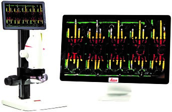
Did coca cola create the modern image of santa. Originally images were captured by photographic film but modern developments in complementary metal oxide semiconductor and charge coupled device ccd cameras allow the capture of digital images fig. Blank one can view a particle under a microscope while a beam of light is directed at the particle in order to obtain its absorption spectrum.
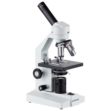
The optical microscope also referred to as a light microscope is a type of microscope that commonly uses visible light and a system of lenses to generate magnified images of small objects. Viruses are too small to be seen directly with a light microscopecan be seen when its examined under an electron. Basic optical microscopes can be very simple although many complex.
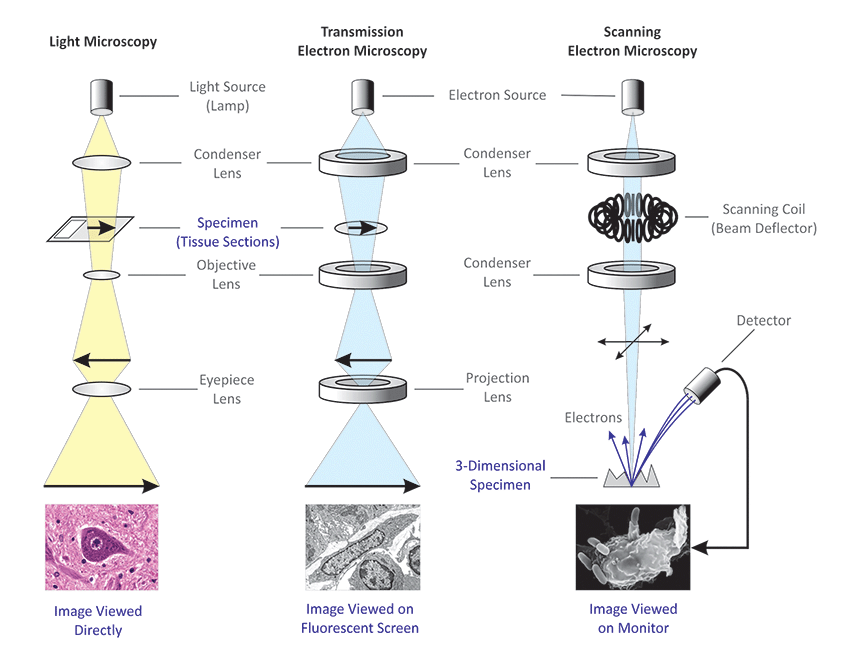
Electron microscopes require use of a fluorescent screen photographic plate or electronic display because electrons cannot be observed directly by the human eye. The electron microscope is a type of microscope that uses a beam of electrons to create an image of the specimen. A type of image that cannot be viewed directly is called a blank image.
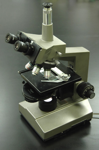
The compound light microscope uses visible light to directly illuminate specimens in a two lens system resulting in the illuminated specimen appearing dark against a bright background. Note how the sem image allows for clear perception of the fine structure details which are hard to fully make out in the light microscope image. Whats the function of the ocular.
Optical microscopes are the oldest design of microscope and were possibly invented in their present compound form in the 17th century. Because the microscope can only focus on one hair at a time. Light microscope images can be viewed directly.
Start studying criminalistics chapter 7. A scanning electron microscope. When scanning objective is in place.
Two images of the same depth hoar snow crystal viewed through a light microscope left and as an sem image right. How is the color distributed when viewed under the microscope patterns. Electron microscopes require use of a fluorescent screen photographic plate or electronic display because electrons cannot be observed directly by the human eye.
 Microscopy Biology For Majors I
 Light Microscopes An Overview Sciencedirect Topics
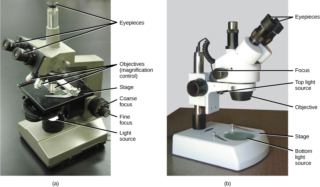
 Instruments Of Microscopy Microbiology
 3 1 As Unit F211 Cells Exchange And Transport The Cell Is The
 How To Use A Microscope With Pictures Wikihow
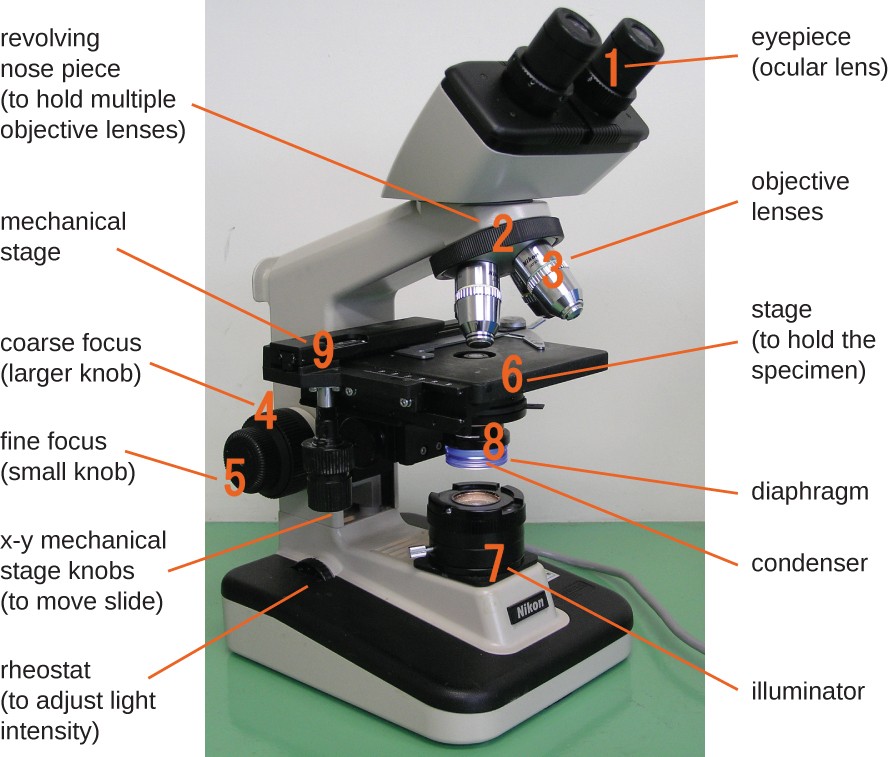
 The Parts Of A Compound Microscope And How To Handle Them
 Optical Microscope An Overview Sciencedirect Topics
 Microscopy Biology For Life
![]() 2018 Hm Transmission Electron Microscope
2018 Hm Transmission Electron Microscope
 Microscopy Intro To Microscopes How They Work Article Khan